Mild traumatic brain injury (TBI), commonly referred to as concussion, describes the acute neurophysiological effects of blunt impact or other mechanical energy applied to the head.1 It is among the most commonly reported neurological conditions, with an estimated annual incidence of 200 out of 100,000 individuals in the United States.2
Physical rest and passage of time remain the traditional primary treatment for acute symptoms of concussion.3 In recent years, the importance of cognitive as well as physical rest has been demonstrated.4 Thus, return-to-activity decisions are often based on a week or more of physical and cognitive rest, with recovery determined by qualitative assessments of patient symptoms.
However, it has long been understood a significant percentage of concussions involve damage to the vestibular system instead of or in addition to the brain. More than 30 years ago, vestibular disorder was objectively detected in 40% and 60% of patients reporting mild and moderate post-traumatic vertigo, respectively.5
UNDERSTANDING VESTIBULAR CONCUSSION AND CEREBRAL CONCUSSION
Periods of rest can actually slow the recovery from a vestibular concussion, causing slower reflex speed, poor balance, and muscular weakness. This factor alone can dramatically affect a patient’s ability and safety to return to activity.
Furthermore, the patient may be at risk of developing post-concussion syndrome (PCS). PCS refers to an array of symptoms resulting from a concussion that does not resolve independently with time. The most common complaints are headaches, dizziness, fatigue, irritability, anxiety, insomnia, loss of consciousness and memory, and noise sensitivity.
The prevalence of PCS in the first weeks after mild TBI varies from 40% to 80%. However, as many as 50% of patients report symptoms for up to 3 months, and 10% to 15% for more than a year.6
Dizziness is the primary symptom indicating vestibular injury. It has been reported to occur in 23% to 81% of cases of concussion in the first days after injury.7 The wide range is likely driven by the vague nature of the term “dizziness.” Dizziness may be perceived in different ways by different individuals. It could be experienced as vertigo, nausea, lightheadedness, or feeling unsteady on one’s feet.
Cerebral concussion and vestibular concussion have similar symptom profiles, and because patient-reported dizziness is subjective, testing is required to conclusively determine the presence of vestibular injury. Physical therapists can play a significant role in the differential diagnosis of
vestibular concussion by providing testing that is typically unavailable through general medical practice.
First, a high-level review of vestibular physiology: Vestibular sensation is mediated by two relatively simple reflexes. The vestibulo-ocular reflex (VOR) ensures optimal visual processing during head motion by stabilizing the line of sight, also called gaze. The vestibulospinal reflexes (VSR) facilitate changes in antigravity muscles to help keep the head and body upright.
There are two sensors within the vestibular labyrinth that respond to acceleration and thereby transduce the motion and position of the head into peripheral and ultimately central biological signals: the semicircular canals (SCCs) and the otolith organs.
The SCCs sense angular acceleration in process information concerning head rotation and head velocity. Otolith organs sense linear acceleration to detect head translation and head position relative to
the gravitational field (ie, uprightness).
In addition to gaze stability (VOR), the SCCs and otoliths provide information for VSR reflexes that keep the head and body upright. These sensors can detect motion in three dimensions, ie, around any or all of the three axes of rotation, pitch (vertical), yaw (horizontal), and roll (torsion), and around any or all of the three axes of translation, fore and aft, side to side, and up and down.
Testing modalities can be divided into three categories: questionnaires, diagnostic, and function.
QUESTIONNAIRES (IE, SELF-REPORTED)
• Dizziness Handicap Inventory (DHI) Score: The DHI measures the perceived level of handicap associated with the symptom of dizziness.8 It includes 25 items with three response levels, subgrouped into three domains: functional, emotional, and physical. It has been used predominantly in patients with peripheral and central vestibular pathology, and to evaluate subjective dizziness impairment in subjects with traumatic brain injury.9
• Activities Specific Balance Confidence (ABC) Score: Questionnaire used to assess respondents’ level of confidence about losing balance while performing 16 functional activities. Highest possible score of 100 suggests maximum confidence and a score of 0 suggests no confidence.10
• Neurocognitive Screen (eg, imPACT): ImPACT records demographic information, administers six neuropsychological tests, and assesses postconcussion symptoms. It then generates composite scores that assess verbal memory, visual memory, reaction time, visual motor processing speed, and impulse control. ImPACT is demonstrated as a valid and reliable measure of neurocognitive deficits following concussion and is currently used to guide decision-making for returning players to sport and safely manage concussions in nonathletes.11-15
DIAGNOSTICS
Several types of diagnostic tests are available to detect the presence of a vestibular component in concussion. Following are the most important available tools:
1. VOR diagnostics:
i. Video nystagmography (VNG) is the mainstay of assessment of the vestibular system in the postconcussed athlete, measuring peripheral and central components of the vestibular system. It is made up of four distinct tests, each able to identify areas of the vestibular system that may be causing persistent symptoms.
ii. Oculomotor testing measures central vestibular function through quantitative measurements of the saccade, smooth pursuit, and optokinetics reflexes. It is recommended that the clinician question the patient to pinpoint whether symptoms are aggravated or reproduced.16
iii. Spontaneous nystagmus testing documents and measures inability of the eyes to maintain a static position as a result of peripheral, CNS, or congenital abnormality. Tests are conducted with eyes open and closed and in “eyes forward,” “eyes right,” and “eyes left” positions.
iv. Positional nystagmus tests measure tone imbalance of the vestibular system, and include the Dix-Hallpike test for benign paroxysmal positional vertigo (BPPV) to determine the involvement of the posterior semicircular canal (P-SCC)17 and the roll test in cases of horizontal semicircular canal (H-SCC) BPPV.18 In the event that the Dix-Hallpike cannot be performed easily, the side-lying testing position may be used.19
v. Caloric testing evaluates the viability of the peripheral end organs by stimulating them with warm and cool water while the patient’s eyes are closed. The resulting dizziness and nystagmus are taken as an index of the viability of the organ. The eyes are then opened to evaluate the ability of the CNS to visually suppress inappropriate dizziness and nystagmus. Water calorics are preferred over air, but measurement allows identification of superior vestibular nerve and/or horizontal canal injury. Air calorics can prove convenient for patient and clinician in certain situations in which water may be contradicted.20 Additional testing alternatives include wet air (WAI), which can be used as an alternative caloric testing method for anxious subjects oversensitive to water testing and potentially for patients with suspected pathological ear canal and tympanic membrane findings.21
vi. Dynamic Visual Acuity Test (DVAT) and Gaze Stabilization Test (GST): The DVAT measures physiology of the VOR, while GST measures function of the VOR.
a. Vestibular Autorotation Test (VAT) allows the clinician to measure the function of the VOR across high frequencies of 2 to 6 Hz to test both horizontal and vertical VOR responses.22
b. Vestibular Evoked Myogenic Potentials (VEMP) measures saccular function and the inferior vestibular nerve.
2. VSR diagnostics:
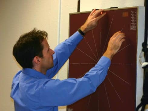
i. Computerized Dynamic Posturography (CDP).
VSR function is measured through various types of computerized posturaltesting. One of the most widely used is CDP, which provides a quantifiable assessment of an individual’s postural control and gait stability. The outcome can determine the functionality of the somatosensory, vestibular, and visual inputs for clinical interpretation.
a. The Sensory Organization Test (SOT) uses a sway referencing of a computerized plate to measure the organization of the balance system.
b. The Sensory Integration Test (SIT) is a new component that measures the function of the balance/vestibular system with a sway randoming.
c. The Head Shake-Sensory Organization Test (HS-SOT) measures postural stability with a controlled head movement mimicking active balance function.
3. Function
Includes: cervical assessment, gait assessment, functional gait assessment (FGA), dynamic gait index (DGI), FUKUDA March Test, and exertional testing. RM
Brian K. Werner, MPT, owns Werner Institute for Balance and Dizziness, Las Vegas. He is Herdman certified (2000) and trained at Northern Arizona University. For more information, contact [email protected].
REFERENCES
1. Bigler ED. Neuropsychology and clinical neuroscience of persistent post-concussive syndrome. J Int Neuropsychol Soc. 2008;14:1-22.
2. Kraus J, Schaffer K, Ayers K, Stenehjem J, Shen K. Physical complaints, medical service use, and social and employment changes following traumatic brain injury: a 6-month longitudinal study. J Head Trauma Rehabil. 2005;20:239-256.
3. Leddy JJ, Sandhu H, Sodhi V, Baker JG, Willer B. Rehabilitation of concussion and post-concussion syndrome. Sports Health. 2012;4(2):147-54.
4. Moser R, Glatts C, Schatz P. Efficacy of immediate and delayed cognitive and physical rest for treatment of sports-related concussion. J Pediatr. 2012;161(5):922-926.
5. Berman J, Fredrickson J. Vertigo after head injury: a five-year follow-up. J Otolaryngol. 1978;7(3):237-245.
6. Spinos P, Sakellaropoulos G, Georgiopoulos M, et al. Postconcussion syndrome after mild traumatic brain injury in Western Greece. J Trauma. 2010;69(4):789-94.
7. Alsalaheen BA, Mucha A, Morris LO, et al. Vestibular rehabilitation for dizziness and balance disorders after concussion. J Neurol Phys Ther. 2010;34:87-93.
8. Jacobson GP, Newman CW. The development of the Dizziness Handicap Inventory. Arch Otolaryngol Head Neck Surg. 1990;116:424–427.
9. Kaufman KR, Brey RH, Chou LS, Rabatin A, Brown AW, Basford JR. Comparison of subjective and objective measurements of balance disorders following traumatic brain injury. Med Eng Phys. 2006;28(3):234-239.
10. Powell LE, Myers AM. The Activities-specific Balance Confidence (ABC) Scale. J Gerontol A Biol Sci Med Sci. 1995;50A:M28–M34.
11. Field M, Maroon J, Collins MW, Lovell MR, Bost J. The use of ImPACT to manage sports-related concussion: Validity data and implications for return to play [abstract]. Neurosurgery. 2003;53:513.
12. Iverson G, Lovell M, Collins M. Validity of ImPACT for measuring the effects of sports-related concussion[abstract]. Arch Clin Neuropsychol. 2002;17:769.
13. Iverson GL, Lovell MR, Collins MW. Interpreting change on ImPACT following sport concussion. Clin Neuropsychol. 2003;17:460-467.
14. Iverson GL, Lovell MR, Collins MW. Validity of ImPACT for measuring processing speed following sports-related concussion. J Clin Experimental Neuropsychol. 2005;27:683-689.
15. Schatz P, Pardini JE, Lovell MR, Collins MW, Podell K. Sensitivity and specificity of the ImPACT test battery for concussion. Arch Clin Neuropsychol. 2006;26:91-99.
16. http://concussion.buffalo.edu/4109Rehabilitation.pdf
17. Korres SG, Balatsouras DG. Diagnostic, pathophysiologic, and therapeutic aspects of benign paroxysmal positional vertigo. Otolaryngol Head Neck Surg. 2004:131(4):438–44. doi:10.1016/j.otohns. 2004.02.046. PMID 15467614.
18. Burak ÖÇ, ?brahim E, Zeynep AÇ, ?enol C, ?brahim S, Suat T. What is the true incidence of horizontal semicircular canal benign paroxysmal positional vertigo? Otolaryngol Head Neck Surg. 2006;134(3):451-454.
19. Cohen HS. Side-lying as an alternative to the Dix-Hallpike test of the posterior canal. Otol Neurotol. 2004;25(2):130-4. PMID 15021771.
20. Zangemeister WH, Bock O. Air versus water caloric test. Clin Otolaryngol Allied Sci. 1980;5:379–387. doi: 10.1111/j.1365-2273.1980.tb02164.x
21. Gudziol H, Koch C, Bitter T, Guntinas-Lichius O. Wet air as an alternative to traditional water irrigation during caloric vestibular testing. Laryngoscope. 2012;122:703–707. doi: 10.1002/lary.22493.
22. O’Leary DP, Davis LL. High-frequency autorotational testing of the vestibulo-ocular reflex. Neurol Clin. 1990;8(2):297-312.


