Understand how to modify detrimental forces at the knee for a rehab program optimized end-to-end

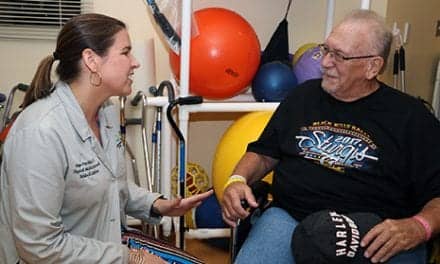
In addition to preparing the patient for surgery, preoperative rehabilitation enables the physical therapist to begin evaluating and correcting specific faulty movement patterns through neuromuscular re-education.
The anterior cruciate ligament (ACL) is the site of the most prevalent knee injury in athletics, with an estimated 200,000 per year or greater in the United States.1 Additionally, females in the athletic population sustain this injury at a rate four to six times greater than their male counterparts in same sport comparisons.2 The goals of this article are to discuss the common detrimental forces at the knee that are modifiable, and their relationship to pre- and post-surgical rehabilitation, and to highlight key patient education opportunities for the therapist during ACL reconstruction (ACLR) rehabilitation management. ACLR protocols and progression milestones are outside the scope of this article.
The role of the ACL is to limit the anterior motion of the tibia and control tibial rotation.3 Injury to the ACL commonly occurs when the anterior shearing force of the tibia under the femur and the torque of that motion are greater than the ACL can withstand. The most common forces to present this load are a combination of forces causing a valgus force at the knee.
ACL injuries occur with both contact and noncontact trauma. With noncontact ACL injury, the valgus loads may be modifiable. Hewett et al has described four neuromuscular imbalances that contribute to noncontact ACL injury in female athletes.4 These imbalances may be amenable to prevention training, and the principles can be used in all patients during preventative, presurgical, and postsurgical rehabilitation. The faulty movement patterns proposed are described below.
- Ligament dominance occurs with insufficient muscle control in the absorption of ground reaction forces that could cause a higher rate of loading to the joint and ligament.
- Quadriceps dominance refers especially to the female athlete’s use of the quadriceps as the primary stabilizer of the knee compared to males who tend to use the posterior chain stabilizers such as gluteals, hamstrings, and gastroc-soleus more efficiently to control loading forces.
- Leg dominance occurs when one leg is favored during tasks that should require symmetrical loading.
- Trunk dominance is described as the inability to precisely sense and control the trunk in three-dimensional space during perturbation and loading.4
Once the quad set, hamstring contraction, and proper pelvis position have been taught on the table, the patient can move to standing to further utilize these patterns.
PREOPERATIVE REHABILITATION
Ideally, physical therapy begins before surgery. Initiating therapy prior to surgery gives the PT the opportunity to initiate patient education and begin to evaluate faulty movement patterns. Preoperative physical therapy also allows the patient to gain confidence in the therapist. Preoperative goals include inflammation management, range of motion (ROM), assessment of correctable movement impairments, and patient education.
Restoring complete passive range of motion preoperatively is a key to the entire rehabilitation program.5 Preoperatively, the priority is to gain normal knee flexion and extension and symmetry between sides.5 Obtaining symmetry allows the therapist to more efficiently determine the real function of the patient’s knee and their readiness for surgery.
Inflammation reduction is imperative in obtaining the goal of symmetrical ROM. Aggressive icing and patient education regarding the importance of having a “calm knee” prior to surgery are key at this time. Instructing the patient to avoid aggravating activities and prolonged positions will assist in reducing irritation in the postinjury/preoperative phase.
In addition to preparing the patient for surgery, preoperative rehabilitation enables the physical therapist to begin evaluating and correcting specific faulty movement patterns through neuromuscular re-education. If the patient’s knee is calm enough prior to surgery, strengthening can be initiated to improve function prior to reconstruction or as a first step in nonoperative management.6 Initiating simple gait training for good movement patterns such as symmetrical load bearing and stance time will assist the patient pre- and post-surgery. Additionally, any of the four factors described above that may have contributed to the injury in the first place can begin to be addressed. These patterns and treatment options will be discussed in the postsurgery section.
Presurgical physical therapy affords time to educate the patient regarding the anatomy and physiology of the postinjury knee as well as to prepare the patient for what to expect after surgery. It is important to address the pain they may be experiencing and explain that while pain is a normal protective mechanism, it does not necessarily define the severity of the injury. Decreasing the threat to the patient is important and often can be accomplished by simply explaining that even in the case of a complete rupture, the ROM exercises will not harm the knee even though they may feel pain.7 This is all within reason, of course, as people have many different experiences of pain and disability. The key point here is that the confidence gained with this knowledge can often be immeasurable and carry a patient through the early challenging phase post-injury and post-surgery. Exposure to specific exercises for range of motion and proper weight bearing is another crucial piece of education that takes place in the presurgical phase. If the patient becomes an expert at certain exercises preoperatively and is educated to begin performing them immediately after surgery, they may gain confidence, control, understandable goals, and realistic expectations.
POSTOPERATIVE REHABILITATION
While the surgeon is focused on strength of graft fixation, reproduction of anatomical footprint, and integrity of graft material, the physical therapist is now focused on ROM, strength, movement patterns, and return to function.8 Early postoperative goals are ROM and inflammation management. To achieve the postsurgical outcome of return to function and sport, it is important to begin to address the modifiable movement patterns as early as possible, beginning while working toward full ROM on the table and progressing with activities as simple as walking.
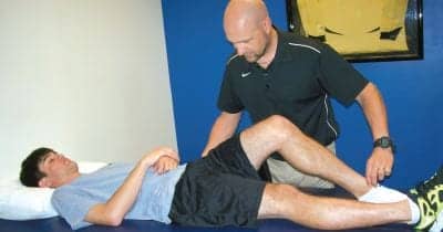
Gaining full active and passive ROM immediately after surgery is associated with decreased risk of postsurgical complications.8,9 A combination of patello-femoral and tibio-femoral mobilizations, retrograde massage, and passive ROM activities are useful at this time in achieving this goal. As passive extension ROM normalizes in this early stage, it is important to cue the patient to activate the quadriceps in what may be new extension range at this time. While the patient is on the table performing quad sets in this new range, the importance of this muscular control can be explained. If tolerated, the quad set should be progressed toward symmetry with the uninvolved leg.5 An added benefit of gaining active terminal knee extension control is swelling reduction through muscle pumping. The patient will eventually be able to activate the quadriceps enough to perform a straight leg raise, enabling progression of weight bearing and gait.
While still supine, passive and active assisted flexion ROM is initiated. Important movement pattern teaching can occur as early as the first day of physical therapy while working toward flexion ROM. Many patients make compensations at the pelvis such as hip hiking as the therapist passively flexes the postsurgical knee. This may be the first opportunity to correct a faulty movement pattern by cueing the patient to maintain a level pelvis. Eventually, the patient can be cued with activation of abdominals, hip flexors, and hamstrings to perform active knee flexion as a supine heel drag. Patient education is key here, and the supine heel drag becomes functional beyond gaining knee flexion ROM in terms of posterior muscle activation. As the patient begins to weight bear and gait train, activation of the posterior structures will help the patient begin to move in patterns that avoid quadriceps dominant and trunk dominant movement patterns.
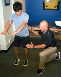
Once the quad set, hamstring contraction, and proper pelvis position have been taught on the table, the patient can move to standing to further utilize these patterns. Teaching the patient to maintain a level pelvis in weight bearing can be initiated by cueing from the top down. If the patient is able to hold the core stable and maintain the pelvis square to the direction of stepping, they can initiate a step by flexing the hip on the involved side with the tibia and foot in neutral rotation.
Very important movement patterns that will carry over into all phases of ACL rehabilitation can be taught as the patient makes contact with the ground at heel strike on the involved side. By demonstration and tactile cueing, the patient can be taught to maintain a neutral pelvis and neutral rotation of femur, tibia, and foot as the heel of the involved side hits the ground. Asking the patient to contract the gluteals and quadriceps as weight shifts forward onto the involved leg promotes greater use of prime movers and posterior chain firing as the involved leg is loaded. This landing/loading pattern will be the key to future progression into squatting, single leg stance, and eventually running, jumping, and landing.
The side weight shift is another simple and important movement pattern to teach. Cueing the patient to weight bear symmetrically with slight hip and knee flexion and the trunk slightly forward over a neutral pelvis loads the posterior structures. The patient is then instructed to slightly shift over the involved leg, keeping the axilla, hip, lateral knee, and lateral malleolus in line in the sagittal plane. This is a subtle shift in weight to teach weight bearing over the involved side that prevents a femoral internal rotation moment and torque on the knee. Cueing the patient to avoid a trunk lean or pelvis rotation while keeping the femur, tibia, and foot in line maximizes the recruitment of gluteals.
In this early postoperative stage, proper cueing of a mini squat is invaluable in addressing and beginning to correct all four faulty movement patterns. Here the initial cue is to break into hip flexion first and allow the weight to shift slightly posterior. As this exercise is progressed, the knees then flex and the trunk and center of mass are cued to move forward over the mid femur, taking care to prevent lumbar extension or anterior pelvic tilting. Symmetric loading of left and right is reinforced as the squat occurs. At the early stage, the emphasis on the timing of hip flexion prior to knee flexion is much more important than the depth of the squat. This movement pattern will be crucial as the patient progresses into higher level activity such as sit to stand, full squatting, and eventually jumping and landing.
The faulty movement patterns described previously are associated with increased risk of ACL injury.4 Once they are identified, therapists can have a significant impact with prevention programs to educate and train patients to avoid these faulty patterns, potentially decreasing noncontact ACL injury rates. In the rehabilitative process, the therapist can use their awareness of these patterns to better educate and treat patients by building a solid foundation for movement starting early after ACL injury and progressing through all phases of postsurgical rehabilitation.
Orr Limpisvasti, MD, is an orthopedic surgeon and sports medicine specialist at Kerlan-Jobe Orthopaedic Clinic in Los Angeles.
Chris Graham, PT, MPT, OCS, is a physical therapist and co-founder of Sports Medicine Institute Los Angeles. He is an orthopedic clinical specialist and has been practicing since 1999.
REFERENCES
- Airelle O Hunter-Giordano, Erin Burlovich, and Tara Jo Manal. Rehabilitation Following Anterior Cruciate Ligament Reconstruction Update of Anterior Cruciate Ligament Injuries. Independent Study Course. 1009 Orthopedic Section, American Physical Therapy Association.
- Arendt EA, Agel J, Dick R. Anterior cruciate ligament injury patterns among collegiate men and women. J Ath Train 1999;2:86-92.
- Markof KL, Burchfield DM, Shapiro MM, Shepard MF, Finerman GAM, Slauterbeck JL. Combined knee loading states that generate high anterior cruciate ligament forces. J Ortho Res 1995;13:930-935.
- Hewett TE, Ford KR, Hoogenboom BJ, Myer GD. Understanding and preventing ACL injuries: current biomechanical and epidemiologic considerations update 2010. N Am J Sports Phys Ther 2010;4:234-251.
- Biggs A, Jenkins WL, Urch SE, Shelbourne KD. Rehabilitation for patients following ACL reconstruction: a value symmetry model. N Am J Sports Phys Ther 2009;1:2-12.
- Eitzen I, Moksaes H, Snyder-Mackler L, Risberg MA. A progressive 5-week exercise therapy program leads to significant improvement in knee function early after anterior cruciate ligament injury. J Orthop Sports Phys Ther 2010;40:705-721.
- Butler, David, and Dr. Lorimer Moseley. Explain Pain. 1st ed.
- Rubinstein RAJ, Shelbourne KD, Van Meter CD, McCarroll JR, Rettig AC, Gloyeske RL. Effect on knee stability after autogenous bone-patellar tendon-bone anterior cruciate ligament reconstruction. Am J Sports Med 1995;23:365-368.
- Mauro CS, Irrgang JJ, Williams BA, Harner C. Loss of extension following anterior cruciate ligament reconstruction: analysis of incidence and etiology using IKDC criteria. Arthroscopy 2008;2:146-153.