
Experienced therapists typically agree that “pain chasing” is the pitfall to solving a complicated case. Therapists cannot simply treat the pain without determining the origin of the pain.
When faced with the task of treating an individual with osteoarthritis (OA), any medical practitioner must begin with the simple question, “What caused the OA in this joint.” Unfortunately, this question is often skipped and symptom management is subsequently initiated. As physical therapists, we run to our comfort zone of electrical stimulation, ultrasound, and—perhaps if we are on the cutting edge—cold laser therapy. The research is fairly scattered regarding the effectiveness of these modalities for pain caused by OA, but anecdotally most therapists will say: “It works for my patients.” The overall results of the studies and systemic reviews in literature demonstrate little to no changes in long-term pain as the result of the use of modalities. However, upon closer inspection, the emerging theme in protocol is to utilize the modality to directly treat the painful joint. This may, in fact, be the disconnect between the literature and actual practice. Experienced therapists typically agree that “pain chasing” is the pitfall to solving the complicated case.
FINDING THE ROOTS OF PAIN
One of the luxuries of being a physical therapist is having the proverbial toolbox of assessment tools and treatment options. When addressing a painful joint, the therapist must first assess the joint, then instantly turn to the joint(s) distal and proximal to that joint. When faced with knee OA, for example, the numerous possibilities of the knee itself must first be addressed; increased Q angle, lack of terminal knee extension, hamstring tightness, and poor quadriceps strength/control, to name a few. But that is just the tip of the iceberg. What about the hip? Does the individual lack full hip extension, limiting full knee extension in standing, thus putting more strain on the already mechanically disadvantaged quadriceps? Or is it the excessive femoral anteversion associated with poor gluteal strength that is increasing the Q angle and placing excessive pressure on the femoral condyles? Now to the ankle/foot complex, is the patient’s pes planus preventing the ability to glide properly at the knee during gait? Or is it the shortened Achilles that is causing excessive recurvatum? These scenarios represent an extremely limited list of possibilities, but the point is clear: therapists cannot simply treat the pain without determining the origin of the pain. Thus, when exploring the topic of modalities for the treatment of OA, we must recognize that the e-stim pads may not always be appropriately placed directly over the knee.
While the history of thermal modalities can arguably be dated back to the dawn of time, historically, advanced modalities have been used for the treatment of pain since 46 AD when Scribonius Largus first used the torpedo fish (electric ray) for the treatment of gout and chronic headaches.1 The science was advanced considerably by Faraday with the discovery of the alternating current in the 1830s, thus opening the door for Melzack and Wall’s pain gate theory in 1965. This was preceded by the use of therapeutic ultrasound in the aftermath of World War II. Since the inception of therapeutic modalities, physical therapists have been at the forefront of their use; however, it was not until recent generations that research has begun to legitimize these uses. Current research and trends have resulted in the use of four main modalities in physical therapy interventions; thermal modalities, ultrasound, electrical stimulation, and low-level laser therapy (LLLT).
THE MAIN PLAYERS
When addressing a painful joint, the therapist must first assess the joint, then instantly turn to the joint(s) distal and proximal to that joint.
Thermal Modalities (Hot and Cold): Thermal modalities are used to reduce pain, speed healing, improve circulation, and increase tissue extensibility. Cold therapy is more useful in the acute phase to reduce inflammation and edema and increase range of motion. Later, heat is useful for increasing flexibility to restore functional mobility when stiffness and soreness set in during the subacute and chronic phases. Studies have shown that for patients with OA, administration of ice massage delivered a clinically important benefit for knee OA, and that cold packs decreased knee edema.2
Ultrasound: Ultrasound is another modality physical therapists can use to generate heat within a body part. This modality uses sound waves rather than external heat. Ultrasound can be used to generate heat in a joint, which will help with overall blood circulation, to loosen up tissues to allow them to respond better to other manual techniques. It also aids in preparing a body part before an exercise program. Ultrasound can be helpful for nonthermal effects as well, by increasing blood flow to reduce inflammation or swelling. A review of the literature reveals that when used on patients with OA, ultrasound has therapeutically beneficial effects on pain, functional outcomes, and cartilage healing properties.3
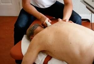
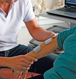
Electrical Stimulation: Electrical stimulation can be used to generate electrical currents for a variety of desired effects. These can include creating muscle contraction to increase strength and prevent disuse atrophy; stimulating muscles, nerves, or sensory nerves to reduce pain; stimulating tissue to speed the healing process of damaged tissue; and preventing scar tissue formation. Electrical stimulation has been found effective for patients with OA in relieving pain and reducing stiffness.4
Low-level Laser Therapy (LLLT): LLLT uses low-level lasers or light-emitting diodes to alter cellular function. Research suggests it is effective in relieving short-term pain for rheumatoid arthritis,5 osteoarthritis,6 and other chronic joint disorders.7.
MAKE THE MOST OF MODALITIES
When treating the cause of the pain rather than the pain location itself, physical therapists can use the potential of the modalities to their fullest. Using therapists’ high level of training to assess and determine the root cause of the pain enables them to take the next step and utilize the technological advances of electrical stimulation, ultrasound, and low-level laser therapy to abolish the pain.
Robert Schreyer, PT, DPT, NCS, MSCS, CSCS, holds a bachelor’s degree from Rutgers University and a doctor of physical therapy degree from New York University. He is a founder and co-owner of Aspire Center for Health & Wellness, and a professor in the Doctor of Physical Therapy Program at Touro College. He also teaches at Columbia University and Hunter College. He is a Neurologic Certified Specialist (NCS) and Multiple Sclerosis Certified Specialist (MSCS). Schreyer has several publications in peer-reviewed journals, has lectured at many conferences, and is co-authoring a chapter for a physical therapy textbook. For more information, contact .
References
- Baldwin B. The Career and Work of Scribonius Largus. Rheinisches Museum für Philologie 135. Frankfurt: Sauerländers Verlag; 1992:74-82.
- Brosseau L, Yonge KA, Robinson V, et al. Thermotherapy for treatment of osteoarthritis. Cochrane Database Syst Rev. 2003;(4):CD004522.
- Srbely JZ, Ultrasound in the management of osteoarthritis: part I: a review of the current literature. J Can Chiropr Assoc. 2008;52(1): 30–37.
- Osiri M, Welch V, Brosseau L, et al. Transcutaneous electrical nerve stimulation for knee osteoarthritis. Cochrane Database Syst Rev. 2000;(4):CD002823.
- Brosseau L, Welch V, Wells GA, et al. Low level laser therapy (Classes I, II and III) in the treatment of rheumatoid arthritis. Cochrane Database Syst Rev. 2000;(2):CD002049. Update 2005;(4).
- Jamtvedt G, Dahm KT, Christie A, et al. Physical therapy interventions for patients with osteoarthritis of the knee: an overview of systematic reviews. Phys Ther. 2007;88:123–136.
- Bjordal JM, Couppé C, Chow RT, Tunér J, Ljunggren EA. A systematic review of low level laser therapy with location-specific doses for pain from chronic joint disorders. Aust J Physiother. 2003;49:107–16.

