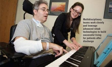
Scientists have shown for the first time that neuroprosthetic brain implants may be able to help stroke patients with partial paralysis.
Researchers have found that implants known as brain-computer interfaces (BCI) may be able to detect activity on one side of the brain that is linked to hand and arm movements on the same side of the body. They hope to use these signals to guide motorized assistance mechanisms that restore mobility in partially paralyzed limbs.
Partial paralysis on one side of the body results from stroke damage to the opposite side of the brain. This fits with the conventional model of how the brain controls movement, which says signals in one half of the brain control the opposite half of the body. That model led scientists to assume that stroke damage would make it impossible for BCIs to pick up any useful movement control signals from the brain and restore function in the body’s paralyzed half.
"In recent years, though, we’ve come to realize that there’s actually some ipsilateral, or same-sided control signals involved in movement," says senior author Eric C. Leuthardt, MD, assistant professor of neurological surgery, neurobiology and biomedical engineering at Washington University School of Medicine in St Louis, and a physician at Barnes-Jewish Hospital. "Now we’ve shown these signals can be detected and are separable from signals that control the opposite side of the body, which means we may be able to use a BCI to restore function."
The study was published online in the journal Stroke.
BCIs bridge gaps from brain damage and other injuries by using implanted electrodes to link the brain to a computer. The implant relays brain signals to the computer, which interprets those signals to control prosthetic devices or other means of interacting with the environment. In an earlier demonstration of the technology’s potential, the same team of scientists showed in 2005 that a patient with a BCI could use the implant to control a video game.
BCIs formerly consisted of small electrodes implanted inside brain tissue to record from individual brain cells. Leuthardt and his colleagues have been developing a different approach known as electrocorticography (ECoG), which uses a plastic sheet filled with electrodes. The sheet rests on the surface of the brain, recording from many neurons at once.
"The old approach was good for acquiring significant signal control, but it suffered from the problem of scar encapsulation," Leuthardt says. "When the electrodes are in the brain for 3 to 6 months, scars will form around them that prohibit them from recording brain signals."
Scar tissue does not form around the ECoG grid because it is implanted on the surface of the brain.
Leuthardt’s team has shown that the ECoG approach can reveal useful insights into what a patient wants to do by analyzing signals from groups of neurons, rather than single neurons. Examples include a desire to move a hand or to speak.
Although the ECoG implants are currently left in place only temporarily, researchers hope to one day implant them for long-term usage.
Wisneski KJ, Anderson N, Schalk G, Smyth M, Moran D, Leuthardt EC. The unique cortical physiology associated with ipsilateral hand movements and neuroprosthetic implications. Stroke, published online Oct.16, 2008
[Source: Newswise]