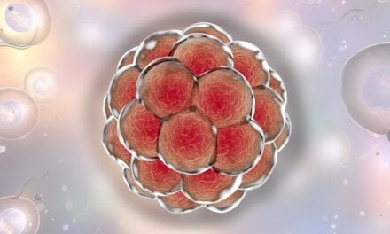
V2a neurons (smaller, faint blue) in the lumbar region of the spinal cord are shown alongside motor neurons (larger, bright blue). (Photo courtesy of Salk Institute)
Research performed on mice has revealed contrasts in the neurons that underlie the motor control of arms and legs. Scientists hope that it could one day lead to tailored stem-cell-based treatments for repairing spinal cord injuries.
“The classic way of thinking about the spinal cord is that it’s a continuous column of neurons that connects to the brain or to the muscles,” says Samuel Pfaff, a Howard Hughes Medical Institute investigator at Salk and senior author of the study, published recently in the journal Neuron.
“If you look at images of spinal cord cross-sections, there might be subtle variations in shape between the areas that control the arms and those that control the legs, but it’s not obvious that there are major differences.”
In the study, the investigators focused on a group of neurons called V2a that express the gene CHX10, explains a media release from Salk Institute.
“We already knew these neurons contribute to movement at all levels of the spinal column,” says Pfaff, who holds the Benjamin H. Lewis Chair. “But this set up a paradoxical situation: What might be different about the V2a neurons at the cervical level, which controls the arms, versus at the lumbar level, which controls the legs?”
Although V2a neurons are present throughout the spinal column, not all of them express CHX10 at the same levels, the researchers observed. First, the team used a technology called RNA sequencing to survey the differences in gene expression of V2a neurons between the arm areas and leg areas (believed to be equivalent to the front and hind limbs in animals).
Marito Hayashi, postdoctoral research associate and first author, discovered that in the mouse spinal cord two major populations of these neurons are graded—transitioning from one population to another. In the cervical (arm) region, the neurons are divided 50-50 between those that express the gene and those that don’t, whereas in the lumbar (leg) region, most of the V2a neurons express it, the release continues.
The investigators used a technology called optogenetics—where light is used to selectively turn on and off cells—to look at how the V2a neurons were connected to muscle-controlling neurons. They found that when V2a neurons were stimulated at the cervical level, the connections to motor neurons were weak, whereas in the lumbar region, the connections were strong and quick.
By next employing a lab technique in which a modified rabies virus is used to trace neural circuitry, the team revealed that in the cervical region, many of the V2a neurons that don’t express Chx10 gene were connected to the brain. By contrast, in the lumbar region where most of the V2a neurons express the gene, the neurons instead were tightly connected to motor neurons and to each other.
This makes sense, according to Pfaff, because hand and arm motions need to be carefully coordinated with the brain, whereas leg movements are more automated.
“Historically, people have treated V2a neurons as one identical population across regions,” Hayashi adds in the release. “But we found that depending on the segment within the spinal cord, their molecular profile—and therefore their job—was different.”
The Salk team, including bioinformatics specialist Shawn Driscoll, looked into whether there were more than two major populations of V2a neurons by employing single-cell RNA sequencing, a technology that allows identification of unique genes expressed at an individual cell resolution. This research led to further identifying 11 unique groups of V2a neurons, per the release.
Future work will focus more closely on the molecular differences between these neurons.
“We hope to do a more detailed analysis to connect different neurons to their functions,” Hayashi concludes.
If their findings also hold true in people, this work could one day lead to tailored treatments for repairing spinal cord injuries, possibly with the use of stem cells, the researchers note.
[Source(s): Salk Institute, Science Daily]