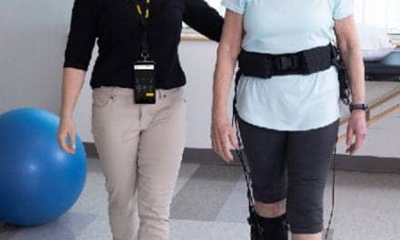

After spinal cord injury, patients want to regain walking ability, but ultimately hand function becomes the top therapy priority.
Following spinal cord injury, patients experience significant disruption in function. For many, the most significant loss is that of hand function. Hand function is compromised with cord injury above the T2 level. This accounts for 47% of the 11,000 new injuries each year.1 Research shows that, while initially patients want to regain walking ability, ultimately they report the loss of hand function as the most debilitating and the first priority for therapy.2 With careful, diligent therapy, patients are able to learn to compensate for loss of function and, in some cases, restore function. Complicating therapy are the cortical changes that occur following injury. The motor cortex shifts, to be biased to rostral to the lesion. Essentially, the brain forgets how to move that which it cannot control. It is essential, therefore, that therapy includes mechanisms for motor learning in addition to the restoration of motor control. Functional electrical stimulation (FES), massed practice, and specific task rehearsal have been shown to yield improvements.3,4
Contracture management and maintenance of the biomechanical balance of the hand should be a primary goal of treatment. In the acute phase, splinting goals should center around maintenance of tendon length. This is most often accomplished by overnight wear of a static resting hand splint, in a neutral or intrinsic-plus position, or with an antispasticity splint, in the presence of hypertonicity. As the patient moves into the subacute phase, static splinting should continue to prevent shortening of soft tissue, especially if tone is an issue, and dynamic splinting is introduced. Dynamic splints are designed to apply a particular torque across a joint to either stretch desired tissues or facilitate a given action. Examples include elbow extension assist splints to prevent biceps shortening, and wrist driven hinge orthoses to facilitate grip. Splints in these categories are generally used long term and may create an aberrant movement pattern through repeated use. It is important, therefore, to evaluate the risks, benefits, and alternatives of these types of splints prior to fabrication. Patient acceptance also should be monitored closely, as these splints can be bulky and difficult to don, leading to poor compliance. In the chronic phase of spinal cord injury, splints are generally used to facilitate a desired function, for example, a wrist cock-up splint with incorporated universal cuff for feeding. At this point, patients have clear preferences regarding function, level of assist, and orthotic management, which should be considered when making recommendations about wear.
From this point, treatment should include a balance of restorative interventions and compensatory training. The compensatory training includes specific task practice, task and environmental modifications, and the inclusion of functional splinting. Restorative interventions aim to provide input to the nervous system that helps to remediate aberrant movement patterns. This often includes massed practice and component level skill training, for example, repetitive grasp and release activities facilitated by FES. There is also evidence to show restorative approaches, ie, near normal levels of input below the level of the lesion, have an excitatory effect on the nervous system, which helps to optimize the nervous system for recovery.5 By including both components, the therapist ensures the patient is able to be independent in the near term, but maintains the potential for more complete recovery in the long term. If treatment focuses solely on compensatory training, or biomechanical changes are allowed to evolve, manifestation of reinnervation may be compromised.
Functional electrical stimulation provides the therapist with a mechanism to move muscles that patients are unable to control on their own. This helps maintain the neuromuscular junction, provides excitatory input to the nervous system, yields a strengthening benefit for emerging reinnervation, and, when coupled with voluntary effort, provides a feedback mechanism for motor learning.3,6 With electrical stimulation, patients are able to reach and grasp, while without electrical stimulation patients may be unable even to lift their arms from a table. There are two major differences in how an FES-driven contraction differs from a physiological contraction. First, since the nerve impulse begins in the middle of the axon, there is not a wave of hyperpolarization behind the impulse. Consequently, the impulse travels retrograde, providing input to and maintaining the motor neuron. This is a desirable consequence. Second, fast twitch motor fibers are preferentially recruited by an electrically driven contraction. This is opposite a physiologically driven contraction, and results in faster fatigue. For this reason, outcomes are best when FES contractions and voluntary contractions can be combined.6,7
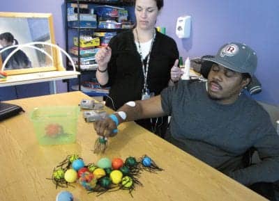
Treating the hand with FES presents a unique set of challenges. There are many small pieces that must fit together in a careful balance to achieve a desired response. This is not easily achieved. The most successful FES applications target gross functions, like grasp and lateral pinch. Repeated training in these patterns has shown some modest improvements in other functions, such as tip pinch and in-hand manipulation. This is primarily attributed to overall strengthening, but is also impacted by changes in the nervous system.8 Using multiple units and triggers allows the therapist to sequence actions and create a more “normal” pattern. In the example pictured, the therapist is using one unit to trigger the triceps to reach and a second unit to sequentially trigger the finger extensors and finger flexors to open and close the hand. Having the patient engaged in the task, even if it is as mundane as moving balls into a container, ensures maximal muscle recruitment and improves motor learning. Motor learning is further enhanced by the afferent input provided by the stimulation and the quantity of repetitive practice.3 A treatment session might include this as a preparatory activity and then transition the same skill into a functional task, such as picking up a cup and drinking from it.
Following is a brief case description that demonstrates how these many pieces fit together.
John is a 21-year-old college senior injured in a motor vehicle accident. He was first diagnosed as having a C5 AIS B injury on the American Spinal Injury Association (ASIA) impairment scale. He was transferred to the International Center for Spinal Cord Injury at Kennedy Krieger Institute for acute rehabilitation 4 months post-injury and spent 5 months as an inpatient at the facility. Initially, John displayed significant tone in his hands, resulting in a fisted hand position that limited his fine motor function. He was treated with Botox to his long finger flexors, sensory level stimulation to modulate tone, and overnight splinting with antispasticity splints. This combination was successful in opening his hands. However, John felt that his hands were too loose and their condition rendered him unable to propel his wheelchair on uneven surfaces and pick up small objects. The splints were modified and the Botox discontinued, allowing for some tightening of the long flexors, but full tendon length has been maintained. In addition to the compensatory training for ADLs, John began a program of FES-assisted grasp-and-release training, which showed modest changes in his hand function.
At discharge from his inpatient admission, John was diagnosed as a C6 AIS C injury on the ASIA impairment scale. He needed supervision for advanced wheelchair skills, moderate assistance for uneven transfers, and moderate assistance for lower body dressing, and was modified independent in all other ADLs including bowel and bladder care.
John returns to our center for outpatient services during school breaks. He feels limited by aberrant movement patterns and finds small item manipulation difficult. Treatment includes specific task practice with the FES-assisted grasp and release training. Additionally, daily FES-assisted upper extremity ergometry has been introduced in outpatient. The ergometry provides patterned stimulation to the nervous system, as does the grasp training, but in a more efficient model and one that John can perform at home. John reports subjective changes, including improved prehension and modulated tone.
The restoration of hand function following spinal cord injury is not an easy task. It involves careful selection of interventions based on the patient’s level of injury, acuity, and personal goals. The treatment requires diligent practice from both the therapist and the patient.
Rebecca Martin, OTR/L, OTD, is the manager of clinical education and training for the International Center for Spinal Cord Injury at the Kennedy Krieger Institute in Baltimore. For more information, visit www.spinalcordrecovery.org.
REFERENCES
- www.sci-info-pages.com/facts.html.
- Ditunno PL, Patrick M, Stineman M, Ditunno JF. Who wants to walk? Preferences for recovery after SCI: a longitudinal and cross sectional study. Spinal Cord. 2008;46:500-506.
- Beekhuizen K, Field-Fote E. Massed practice versus massed practice with stimulation: effects on upper extremity function and cortical plasticity in individuals with incomplete cervical spinal cord injury. Neurorehabilitation and Neural Repair. 2005;19:33-45.
- Mikulis D, Jurkiewicz MT, McIlroy WE, et al. Adaptation in the motor cortex following cervical spinal cord injury. Neurology. 2002;58:794-801.
- Sadowsky CL, McDonald JW. Activity-based restorative therapies: concepts and applications in spinal cord injury-related neurorehabilitation. Devel Disabil Res Rev. 2009;15:112-116.
- Santos M, Zahner LH, McKiernan BJ, Mahnken JD, Quaney B. Neuromuscular electrical stimulation improves severe hand dysfunction for individuals with chronic stroke: a pilot study. J Neurologic Phys Ther. 2006;30:175-183.
- Gordon T, Mao J. Muscle atrophy and procedures for training after spinal cord injury. Phys Ther. 1994;74:56-66.
- Shields RK, Dudley-Javoroski S. Musculoskeletal plasticity after acute spinal cord injury: effects of long-term neuromuscular electrical stimulation training. J Neurophysiol. 2006;95:2380–2390.