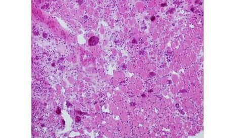
Muscle fibers are shown in pink. The lighter spaces between the fibers indicate defects in muscle regeneration. (Photo courtesy of Institut Pasteur.)
In a recent paper, French scientists suggests that the muscle impairment caused by sepsis may be caused by a drop in cells’ mitochondrial mass.
In the study, published recently in Nature Communications, the researchers also suggest exploring the possibility of using mesenchymal stem cell transplantation as a potential avenue for therapy.
A media release from Institut Pasteur explains that, to help shed further light on the significant loss of muscle capacity observed in patients long-term after recovery, researchers from the Institut Pasteur’s Human Histopathology and Animal Models Unit, directed by Professor Fabrice Chrétien, working together with the research group led by Miria Ricchetti in the Stem Cells and Development Unit (Institut Pasteur/CNRS), investigated the consequences of sepsis on muscle stem cells—known as satellite cells—which develop into muscles, particularly in the limbs. They observed a drastic fall in the mitochondrial mass of these stem cells in mice.
The scientists demonstrated that following sepsis, the few remaining mitochondria provide satellite cells with just enough energy for their basic survival, but not enough for dividing and differentiating into muscle cells when required for muscle growth, repair and maintenance. This damage, which occurs at an early stage and has a long-term impact, prevents the organism from fully restoring muscle function, hence the persistent muscle impairment observed in patients, the release continues.
This research led the scientists to explore the use of mesenchymal stem cells as a possible therapy.
Using a mouse model, Chrétien and his team demonstrated that an intramuscular mesenchymal stem cell transplant carried out after septic shock resulted in a drop in the level of overall inflammation and related symptoms, such as fever, atony (loss of muscle tone), circulation of cytokines, and inflammatory molecules. A histological analysis performed after the transplant showed that the mesenchymal stem cells provided support for the damaged satellite cells without actually replacing them. The mesenchymal stem cells were then eliminated by the organism, with the transplant having successfully repaired the mitochondrial dysfunction and fully restoring the satellite cells’ metabolic and division capability.
[Source(s): Institut Pasteur, EurekAlert]