How newer technologies can help drive motor recovery of upper extremities in stroke patients.
By Megan Palmer, OTR/L
The magic of neuroplasticity following a stroke has long been a phenomenon of neurological recovery. Thousands of studies have been performed to understand specific treatment interventions that drive this neurological change. How is it that damaged or dead brain tissue following a stroke can be circumvented to allow for new nerve pathways that result in motor recovery?
For a considerable time, stroke was known as a focal disease at the area of infarct, but the neurological deficits that present themselves tend to have an impact on areas distal to the damaged brain tissue, if not to the entire brain. The localized stroke may result in deficits affecting all areas of function including motor, sensory, visual, cognitive, speech, and emotional.
A neuroimaging technique called diffusion weighted imaging (DWI) provides the ability to analyze in vivo white matter bundles and show how areas of the brain are interconnected. DWI can provide a map of routes for reorganization of neural signals/impulses that can be a highway for neuroplasticity.1
With the information that associated centers of the brain can be simultaneously affected by an infarct in a distal area, neuroscientists strive to identify how treatment interventions related to stroke can be utilized to drive neurological change. The use of Functional Magnetic Resonance Imaging (fMRI), in addition to other neuroimaging technologies, can provide distinctive information on the networking of the human brain and what areas are “lighting up” when certain motor activities are performed. Essentially, fMRI is able to identify areas of increased blood flow associated with active neurons requiring an increase in oxygen supply. This connection can be interpreted as the brain adapting to discover new ways to recover normal motor function.2
Guiding Principles in Motor Recovery
The most common deficit after stroke is hemiparesis of the upper extremity (UE), contralateral to the site of the brain infarct. Eighty-five percent of stroke survivors experience hemiparesis and 65% result in functional limitations. These are striking numbers that directly impact a patient’s quality of life and determine his or her level of dependence on others.3
Utilizing the methodology that neural recovery is possible via fMRI data can guide therapeutic interventions ensuring high-quality, high dosage, high repetitions of normal movement patterns to aid in the motor recovery of the hemiplegic UE.Research has shown there is no specific prescription or protocol for rehabilitation after stroke, but there are principles to guide current practice:
• Specificity—Need to be direct with treatment intervention to enhance neuroplasticity; task-specific training.
• Repetition—Repeat movement patterns; practice in the way that is going to be effective for the rest of their lives (do not teach compensatory movements with the affected UE).
• Intensity—It matters; work until failure; take to the point that they cannot perform the activity anymore; push them, challenge them; the demand is what is going to drive change in the brain.
• Salience—Has to mean something to the patient, they have to buy-in, physiological changes in the brain are to a greater degree when activity is purposeful.
• Allow for patient error—Neuroplastic changes occur in the midst of errors, error-correction, failure, near-misses, and exploration.
• Utilize an enriched environment—Integrate sensory and intellectual stimulation.
Neurological change occurs when you practice the next level, add variables, and provide the just-right challenge for the patient. This rewiring of the brain does not occur in stagnant environments, with the practice of already mastered skills or with familiar tasks. Treatment interventions need to be tailored to the patient’s goals and relate to movements that carry over to functional tasks.4
Due to the variability of stroke paralysis that occurs in each patient and the limiting factors including spasticity and tone, it is stated that a variety of conventional methods and technologies should be used to treat hemiplegia post-stroke. Additional technologies have proven to be an effective adjunct to conventional stroke rehabilitation interventions. Electrical stimulation, functional rehabilitation systems, myoelectric devices, and virtual reality are all examples of technologies that aid in stroke rehabilitation of the upper extremity.5
Neuromuscular Electrical Stimulation (NMES) in Motor Recovery
NMES is used to target specific muscle groups for patients with hemiplegia following stroke and promote motor facilitation. It provides an alternate route to gain maximum repetitions of an otherwise paralyzed muscle.
NMES utilizes an electric current that produces a contraction of a muscle with intact lower motor neuron connections. The NMES device allows for adjustments to be made with frequency, pulse width and amplitude that produce a waveform resulting in a visible muscle contraction. The variability of electrode placement and multiple leads allow for the ability to stimulate more than one muscle group at a time. This can assist with providing adequate contractions in the shoulder muscles to decrease subluxation and distal muscles of the upper extremity to produce repetitive stimulation.
Research is inconclusive on exactly how effective electrical stimulation is to recovery of a hemiparetic upper extremity, but it is one of the most common and favorably mentioned in stroke rehabilitation practices.6
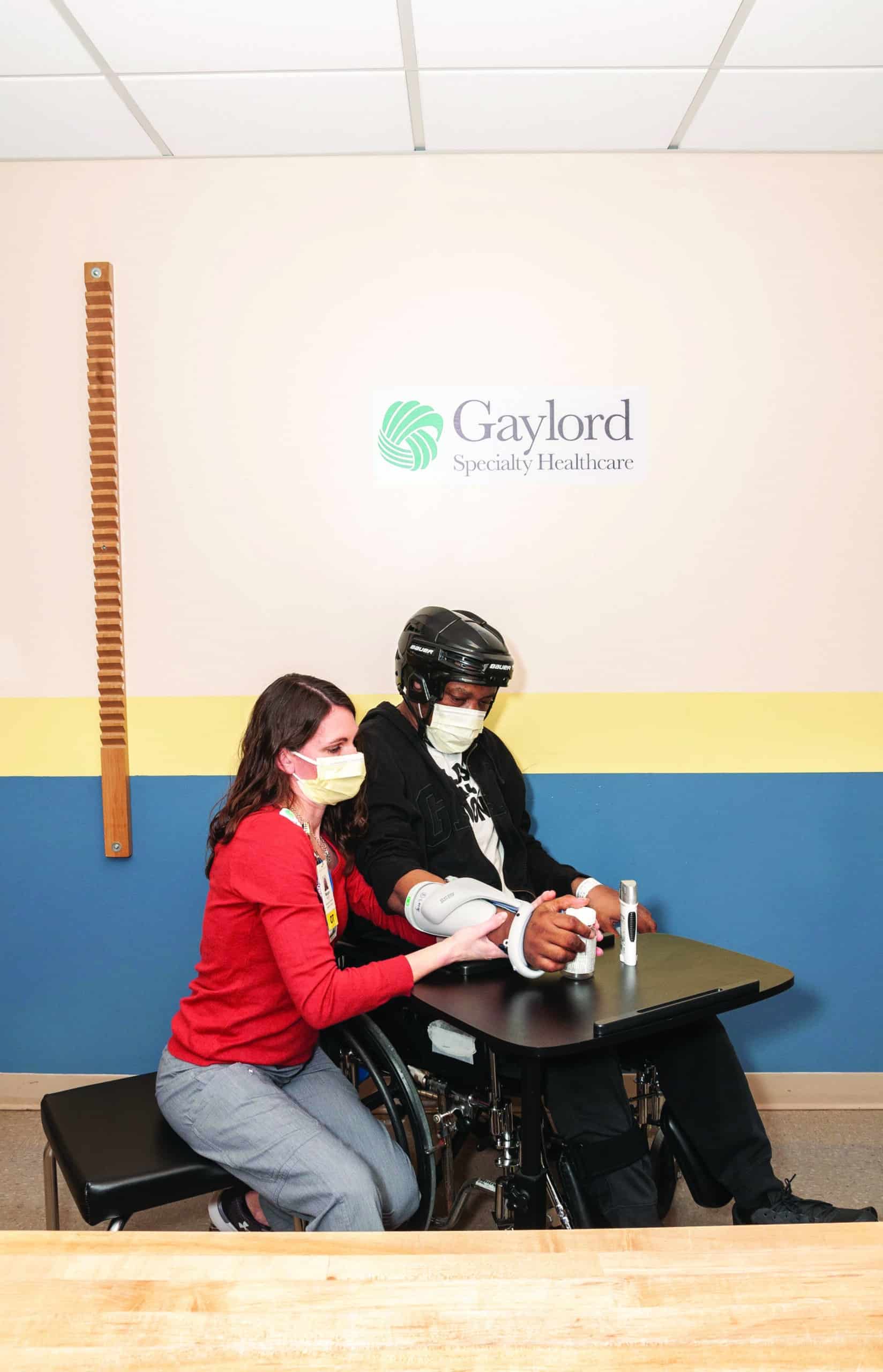
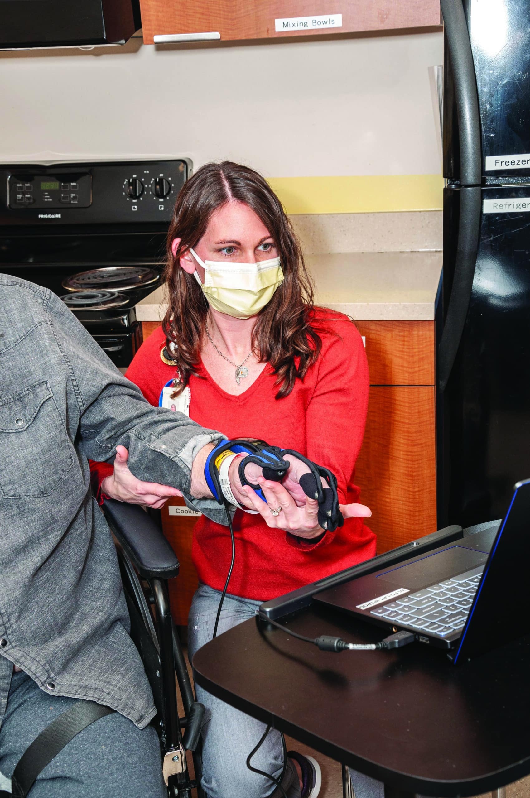
Functional Electrical Stimulation Unit for the Hand
A wireless hand rehabilitation system that provides electrical stimulation to the wrist and digit flexors and extensors is a device that is often used by inpatient and outpatient therapists at Gaylord Specialty Healthcare, where I work.
The device is applied to the forearm and has five electrodes that stimulate the extensor digitorum, extensor pollicis brevis, flexor digitorum superficialis, flexor pollicis longus, and thenar muscles to promote gross grasp and release.7 The unit allows the UE to be incorporated into a functional grasp/release task such as holding a cup and bringing it to the mouth or holding a jar while the unaffected UE opens it. These functions can open a world of opportunity for a patient with hemiparesis if used in their daily life. Due to the repetitive nature and neuromuscular stimulation, this technology also has been favored to aid in neurological recovery.
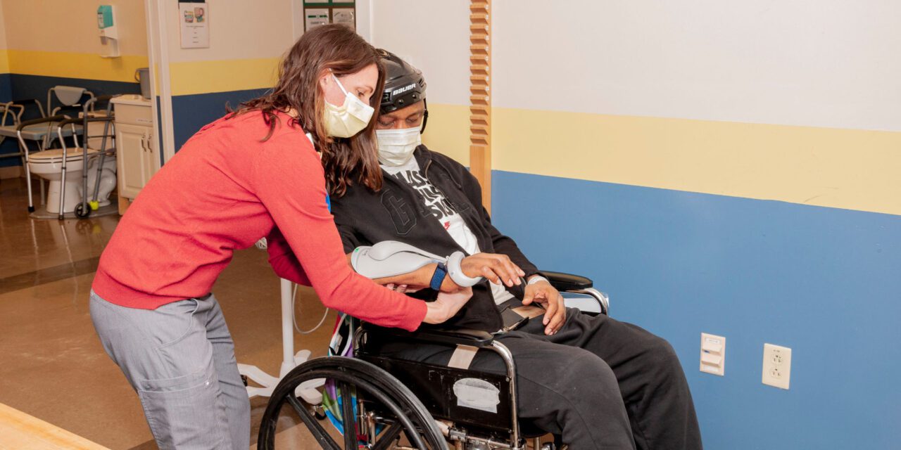
Myoelectric Upper Limb Orthoses for Stroke Rehab
A neuro-robotic arm brace was designed to assist in motor recovery of the upper extremity following a stroke. Devices like this fit over the elbow and secure on the proximal humerus and distal forearm. Sensors are built into the cuffs that align over the biceps and triceps to detect electromyographic (EMG) signals when the patient initiates movement into elbow flexion or extension. This triggers the device to assist in completing the full range of motion in that particular plane. This allows repetitive biceps/triceps movement related to function that assists in rebuilding the neural pathways to regain control of the affected arm.8
There are also devices adapted from this model with two additional sensors on the forearm to sense an initiation of wrist/hand flexors and extensors to promote multi-joint functional movements. These newer devices were designed for daily use and are lightweight for patients’ comfort while performing daily activities or job tasks.9 Studies have shown that the use of portable, myoelectric elbow-wrist-hand orthoses have promising effects on improved motor recovery of the upper extremity, but are vague on reporting the long-term lasting effects of motor return when the device is taken off.10
Arm and Hand Rehabilitation Systems
Functional rehabilitation systems for the upper extremities utilize movement biofeedback and the patient’s current range of motion and strength to enhance and drive neurological change.
Gaylord recently invested in a device of this nature combined with virtual gaming to promote neurological return of the upper extremity. In the initial trial period, patients have shown positive engagement in the gaming device when otherwise their participation was minimal. Their attention is focused on the game instead of on the actual number of repetitions they are performing.
The device is placed on the affected UE with the hinge lock at the lateral elbow. The software assesses patients’ active range of motion into elbow flexion and extension and utilizes those movements to promote repetitive exercise in a fun way.
A hand rehabilitation system is the same concept as the arm device, but instead an ergonomic wearable glove is applied to the patient’s affected hand to utilize their current active hand and wrist motion and capitalize on it.11
Virtual Reality
While research is limited on the effects of virtual reality technology improving hemiplegia in stroke patients, we know that it provides an incentive to perform a repetitive exercise when utilizing a gaming system. In the Sevgi, et al12 study and the Kim13 study that examine stroke patients in a control group receiving conventional therapy and an experimental group receiving conventional and use of video games, improvements of UE function were significantly higher in a few of the UE assessments. In fact, the Kim study abstract states that “Stroke patients who completed the additional training using virtual reality games showed significantly greater improvement in their daily living activities than those who only received traditional rehabilitation therapy.”13 According to a study by Kiper et al,14 reinforced feedback in virtual environment (RFVE) therapy combined with conventional rehabilitation treatment promotes better outcomes for upper extremities than the same amount of conventional rehabilitation, for all types of stroke.
Conclusion
In summary, the majority of studies involving stroke rehabilitation technologies state that they may have positive effects on UE function in stroke patients, but additional studies are needed with greater sample sizes and more precise treatment prescriptions. Specifics are needed to identify how much and how long to utilize the technologies to make the greatest significant differences in UE recovery. In addition, the majority of studies do not utilize patients in their first six months post-stroke. Typically, the greatest neurological recovery is seen in the first three-to-six months following a stroke, which would be the ideal time to investigate the effects of conventional versus technological interventions.
In conclusion, review of the studies mentioned with the use of these technologies, including virtual reality, do not show any negative effects; they only contribute to the patient’s motor recovery and function of UE hemiparesis and would overall complement stroke rehabilitation. RM
Megan Palmer, OTR/L, is a Level III inpatient occupational therapist for Gaylord Specialty Healthcare in Wallingford, Conn. Palmer received her Bachelor of Rehabilitation and Disabilities Studies with a minor in Psychology and her Master’s Degree in Occupational Therapy from Springfield College and has been working with patients with traumatic brain injury and stroke for 15 years.
References
1 Guggisberg AG, Koch PJ, Hummel FC, Buetefisch CM. Brain networks and their relevance for stroke rehabilitation. Clin Neurophysiol. 2019;130(7):1098-1124. doi: 10.1016/j.clinph.2019.04.004
2 Crofts A, Kelly ME, Gibson CL. Imaging Functional Recovery Following Ischemic Stroke: Clinical and Preclinical fMRI Studies. J Neuroimaging. 2020;30(1):5-14. doi: 10.1111/jon.12668
3 Gurbuz N, Afsar SI, Ayaş S, Cosar SN. Effect of mirror therapy on upper extremity motor function in stroke patients: a randomized controlled trial. J Phys Ther Sci. 2016;28(9):2501-2506. doi: 10.1589/jpts.28.2501
4 Green, M. (Aug 2021). PESI, Inc. Stroke Rehab Master Class – Best Practices for Improved Outcomes. Igniting Neuroplasticity after Stroke: Breakthroughs for Improving Motor Recovery.
5 Ward NS, Brander F, Kelly K. Intensive upper limb neurorehabilitation in chronic stroke: outcomes from the Queen Square programme. Journal of Neurology, Neurosurgery & Psychiatry. 2019;90(5):498-506.
6 Knutson JS, Fu MJ, Sheffler LR, Chae J. Neuromuscular Electrical Stimulation for Motor Restoration in Hemiplegia. Phys Med Rehabil Clin N Am. 2015;26(4):729-745. doi: 10.1016/j.pmr.2015.06.002
7 Takeda K, Tanino G, Miyasaka H. Review of devices used in neuromuscular electrical stimulation for stroke rehabilitation. Med Devices (Auckl). 2017;10:207-213. Published 2017 Aug 24. doi: 10.2147/MDER.S123464
8 Wasielewski D. 2010, April. Myomo Robotic Therapy Device. StrokeNetwork Newsletter. Retrieved from Stroke Network: http://strokenetwork.org/newsletter/therapies/myomo.htm
9 Demaitre E. (2019, June 7). Myomo scales up production, training for MyoPro device for upper-body mobility.
10 Peters HT, Page SJ, Persch A. Giving Them a Hand: Wearing a Myoelectric Elbow-Wrist-Hand Orthosis Reduces Upper Extremity Impairment in Chronic Stroke. Arch Phys Med Rehabil. 2017;98(9):1821-1827. doi: 10.1016/j.apmr.2016.12.016
11 MediTouch. (2018). MediTouch HandTutor. Retrieved from MediTouch Homepage: https://meditouch.co.il/products/handtutor/
12 Ikbali Afsar S, Mirzayev I, Umit Yemisci O, Cosar Saracgil SN. Virtual Reality in Upper Extremity Rehabilitation of Stroke Patients: A Randomized Controlled Trial. J Stroke Cerebrovasc Dis. 2018;27(12):3473-3478. doi: 10.1016/j.jstrokecerebrovasdis.2018.08.007
13 Kim JH. Effects of a virtual reality video game exercise program on upper extremity function and daily living activities in stroke patients. J Phys Ther Sci. 2018;30(12):1408-1411. doi: 10.1589/jpts.30.1408
14 Kiper P, Szczudlik A, Agostini M, et al. Virtual Reality for Upper Limb Rehabilitation in Subacute and Chronic Stroke: A Randomized Controlled Trial. Arch Phys Med Rehabil. 2018;99(5):834-842.e4. doi: 10.1016/j.apmr.2018.01.023