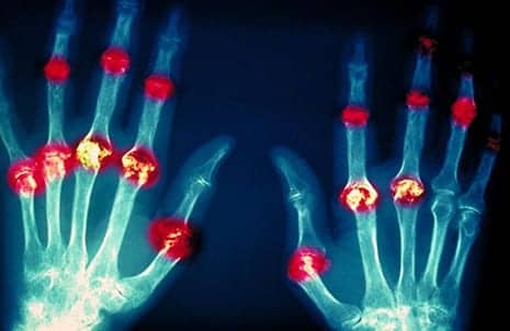
Pictured here is a horizontal x-ray image depicting joint damage from rheumatoid arthritis. (Photo courtesy of Camazine Scott)
Research supported by the Accelerating Medicines Partnership (AMP) on Rheumatoid Arthritis and Systemic Lupus Erythematosus (RA/SLE) provides new insights into tissue damage for these autoimmune conditions and set the stage for uncovering potential drug target candidates that could advance to experimental treatments.
Findings include the identification of novel molecular signatures related to immune system signaling in kidney cells that may reflect their active role in disease process; molecular targets, including specific white blood cells, for potential treatment in lupus nephritis; and specific types of fibroblasts and white blood cells that are involved in rheumatoid arthritis.
Results of the studies were published recently in three papers in Nature Immunology.
A primary goal of the AMP RA/SLE program, which is led by the National Institute of Arthritis and Musculoskeletal and Skin Diseases (NIAMS), is to study tissues where the disease is active in patients, whereas most previous work studied mouse models or only blood samples from humans.
AMP researchers looked at all the cell types in either biopsy samples from kidneys of people with SLE or the synovial tissues of joints from people with RA. The program seeks to quickly find the most promising treatment targets so less time is lost chasing unsuccessful leads, a media release from NIH/National Institute of Arthritis and Musculoskeletal and Skin Diseases explains.
Highlights from the papers, the release continues:
Profiling kidney and skin cells in lupus nephritis. Lupus nephritis is a potentially fatal kidney disease that occurs in about 50% of people with lupus. There can be a wide variety of changes in the kidneys, making the disease hard to diagnose and treat. AMP investigators led by co-senior investigator Jill Buyon, MD, at New York University analyzed a large number of individual cells from kidney and skin samples from people with lupus in order to understand more about the complex mechanisms involved in tissue damage.
Researchers discovered molecular signatures, related to immune system signaling and scar-forming gene activity, in kidney cells that may reflect their active role in disease process. This finding was unexpected since inflammatory cells were thought to be the primary cause of tissue damage. Single cell analysis of skin revealed similar changes, suggesting that in the future it may be possible to monitor a person’s disease progress and treatment responses from skin samples instead of more invasive kidney biopsies.
Understanding the role of immune cells in lupus nephritis. AMP investigators led by Betty Diamond, MD, at The Feinstein Institute for Medical Research, Manhasset, New York, analyzed kidney, blood and urine samples from people with and without lupus nephritis to learn more about how immune cells cause progressive damage in the kidneys.
The scientists uncovered subsets of white blood cells that are active in the disease process and identified molecules that may be potential therapeutic targets. Single cell analysis of immune cells in urine yielded similar results. These findings suggest it may be possible to track immune cell status in the kidneys easily through urine analysis.
Defining inflammatory cell states in rheumatoid arthritis joint tissue. The autoimmune disease rheumatoid arthritis is characterized by chronic inflammation of the synovium – a thin tissue that lines joints. AMP investigators led by Soumya Raychaudhuri, MD, PhD, at Brigham and Women’s Hospital, Boston, used single cell profiling technologies to analyze synovial biopsies.
They identified subsets of cells – including fibroblasts, which are involved in producing cellular scaffolding, specific white blood cells and others – that appear more often in people with rheumatoid arthritis. Researchers will need to determine whether the identified cell subsets are involved in tissue inflammation, and whether targeting these cells could potentially provide a therapeutic benefit. The white blood cells identified may also play a role in other immune diseases.
“These AMP rheumatoid arthritis and lupus findings offer insights into intriguing immune system targets that are worthy of more investigation,” says Robert Carter, MD, acting director of NIAMS.
“We look forward to bringing the most promising of these findings forward to clinical trials, potentially leading to much-needed new treatment options for those living with rheumatoid arthritis, lupus and other immune system disorders.”
To date, the program has made major advances in creating standardized ways to collect kidney and synovial tissue for research in the United States. This standardization has allowed scientists to use state-of-the-art technologies to analyze individual immune cells and other cells from affected tissues. By studying genes, proteins and biological pathways at such high resolution, scientists hoped to uncover novel insights into the mechanisms behind RA and lupus nephritis, a serious complication of lupus.
[Source(s): NIH/National Institute of Arthritis and Musculoskeletal and Skin Diseases, EurekAlert]

